AlphaFold and Protein Folding: Biology's 'Transistor' Moment?
From Levinthal's Paradox to Evoformer and Diffusion architectures, pLDDT/PAE metrics, and de novo enzyme design: A technical analysis of the structural revolution sparked by AlphaFold.
Greetings. In one of our foundational technical analyses at Tech&Lab, “The Cornerstone of the Digital Universe: Transistor Wars”, we examined how our modern digital civilization was erected upon microscopic silicon switches tens of thousands of times thinner than a human hair. Inside an Apple M4 processor, for instance, 28 billion transistors toggle trillions of times per second to execute the logic gates of the digital realm.
What, then, are the “transistors” of the carbon-based biological universe?
The invisible engines sustaining the biochemical machinery of life are proteins. Yet to a bioengineer, a protein is never merely a one-dimensional string of letters (M-A-L-W-M-R...) printed on a page; it is a three-dimensional nanoscale machine that folds with sub-angström precision, alters its conformation, and shuttles electrons and protons. For half a century, the primary bottleneck in molecular biology was calculating how that one-dimensional genetic tape translates into functional three-dimensional mechanical architecture.
Today, structural biology is undergoing a historical inflection point through AlphaFold—strikingly analogous to how the semiconductor industry broke through physical scaling barriers by evolving from 2D Planar FETs to 3D FinFET and GAAFET nanosheet architectures.
1. Introduction: The Universe Where Form Dictates Function
The central dogma of molecular biology dictates that information flows from $\text{DNA} \rightarrow \text{RNA} \rightarrow \text{Protein}$. A newly synthesized polypeptide chain exiting the ribosome is a one-dimensional (1D) linear sequence of 20 standard amino acids linked by peptide bonds (primary structure). The moment this chain enters the aqueous cytosolic environment, however, it folds within milliseconds into alpha-helices ($\alpha$-helices) and beta-sheets ($\beta$-sheets), ultimately locking into an idiosyncratic three-dimensional (3D) topology (tertiary and quaternary structure).
The core axiom of bioengineering is simple: Sequence determines structure, and structure dictates function.
In the 1960s, Christian Anfinsen’s landmark denaturation and refolding experiments on ribonuclease A (Anfinsen’s Thermodynamic Hypothesis) proved that the native 3D conformation of a globular protein under physiological conditions is encoded directly in its amino acid sequence, corresponding to the global free energy minimum ($\Delta G$) of the system:
\[\Delta G = \Delta H - T\Delta S < 0\]An enzyme’s catalytic active site, an antibody’s complementarity-determining regions (CDRs), or the selectivity filter of a transmembrane ion channel are all spatial geometries defined at the angström scale ($1\text{ \AA} = 10^{-10}\text{ m}$).
The Biological Cost of Misfolding
Just as a dielectric breakdown in a single gate oxide produces leakage current that bricks an integrated circuit, protein misfolding shatters cellular homeostasis.
When hydrophobic amino acid side chains that should be buried inside the protein core are erroneously exposed to the cytosol, a thermodynamically unstable state arises. These hydrophobic patches aggregate into insoluble, protease-resistant cross-beta ($\beta$-sheet) amyloid fibrils:
- Alzheimer’s Disease: Extracellular amyloid-$\beta$ ($\text{A}\beta_{42}$) plaques and intracellular neurofibrillary tangles of hyperphosphorylated Tau proteins.
- Prion Diseases (Creutzfeldt-Jakob, BSE): The normal cellular prion protein ($\text{PrP}^\text{C}$, predominantly $\alpha$-helical) converts into the infectious, misfolded isoform ($\text{PrP}^\text{Sc}$, predominantly $\beta$-sheet). Most strikingly, $\text{PrP}^\text{Sc}$ acts as an autocatalytic conformational template, forcing healthy $\text{PrP}^\text{C}$ molecules into its pathological geometry.
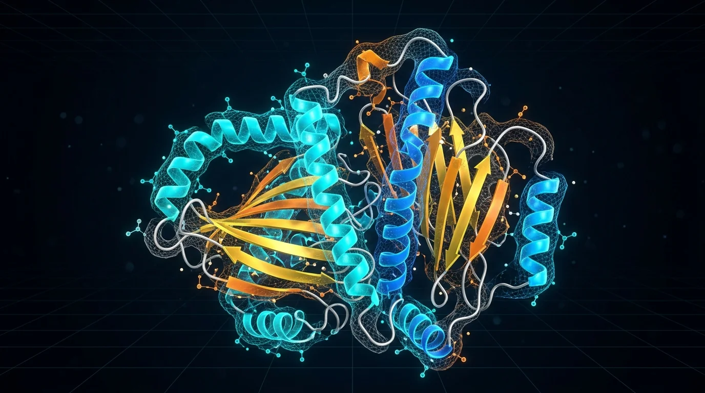 Figure: The thermodynamic trajectory from an unfolded polypeptide chain to a functional native state or pathological amyloid aggregates.
2. Levinthal’s Paradox: Navigating $10^{300}$ Possibilities
If the 1D amino acid sequence deterministically dictates 3D geometry, why couldn’t we solve every protein structure by brute-force numerical integration of quantum mechanical and molecular dynamics equations?
The answer was formulated in 1969 by molecular biologist Cyrus Levinthal.
In a polypeptide backbone, the peptide bond ($\text{C}-\text{N}$, dihedral angle $\omega$) possesses partial double-bond character and is planar and rigid ($\omega \approx 180^\circ$ or $0^\circ$). However, each residue’s alpha-carbon ($\text{C}_\alpha$) possesses two degrees of rotational freedom:
- $\phi$ (Phi): Rotation around the $\text{N}-\text{C}_\alpha$ bond.
- $\psi$ (Psi): Rotation around the $\text{C}_\alpha-\text{C}$ bond.
Additionally, side chains exhibit rotameric dihedral angles ($\chi_1, \chi_2, \dots$). Consider a modest protein composed of $N = 100$ to $300$ residues:
- Even under a coarse, discrete model where each bond assumes only $3$ stable states, a tiny 100-residue peptide exhibits $3^{198} \approx 10^{94}$ backbone conformations.
- Under realistic angular resolutions and side-chain rotamers, the conformational search space explodes to roughly $10^{300}$ states (for perspective, the observable universe contains approximately $10^{80}$ atoms).
Suppose a protein samples these conformations at the rate of atomic vibrational frequencies ($\tau \approx 10^{-13}\text{ s}$, or $10^{13}$ conformations per second). Searching merely $10^{100}$ states would take:
\[t_{\text{search}} = \frac{10^{100}\text{ conformations}}{10^{13}\text{ states/s}} = 10^{87}\text{ seconds} \approx 3.17 \times 10^{79}\text{ years}\]Yet the universe is only $\approx 13.8 \times 10^9\text{ years}$ old ($\approx 4.35 \times 10^{17}\text{ seconds}$).
This is Levinthal’s Paradox: How does an unfolded protein emerging from a ribosome resolve an optimization problem that would take longer than the age of the universe in microseconds ($\mu\text{s}$) to milliseconds ($\text{ms}$)?
Nature’s Solution: The Folding Funnel
Nature does not perform an unbiased random search. Biophysicists model this process as a multidimensional Folding Funnel energy landscape. The wide rim represents high conformational entropy ($\Delta S$) and high free energy, while the narrow bottom corresponds to the singular native state.
As the chain synthesizes, local interactions (hydrophobic collapse, local hydrogen bonding, salt bridges) pull the polypeptide down local thermodynamic energy gradients. Much like Field Marshal Helmuth von Moltke’s strategic doctrine, the initial physical setup (Aufmarsch) is deterministic, but the folding operation proceeds as a stochastic “system of expedients” (System der Aushülfen) dynamically adapting to microscopic fluctuations to funnel into the lowest free energy state.
3. The Limits of Empirical Methods: The Wet-Lab Bottleneck
Prior to the algorithmic breakthrough, resolving the 3D atomic coordinates of a protein ($x, y, z$ coordinates in PDB format) required laborious, expensive, and temperamental experimental techniques:
1. X-Ray Crystallography
The historical gold standard, burdened by a severe engineering bottleneck: Protein crystallization.
- You must recombinantly express the protein at high purity, purify it through affinity and size-exclusion chromatography (SEC), and coax millions of molecules into an ordered, periodic 3D crystal lattice.
- Many critical proteins—such as membrane proteins (GPCRs, ion channels) that constitute over 50% of all modern drug targets—naturally resist crystallization due to flexible loop domains and hydrophobic surfaces.
- Even when a crystal is obtained, X-ray diffraction detectors measure only wave amplitudes ($I_{hkl} \propto \vert{}F_{hkl}\vert{}^2$); the phase angle ($\alpha_{hkl}$) required to compute the electron density map $\rho(x,y,z)$ via inverse Fourier transform is lost (The Phase Problem):
2. Cryogenic Electron Microscopy (Cryo-EM)
Cryo-EM eliminated the crystallization requirement by flash-freezing protein solutions in liquid ethane (vitrification) and reconstructing 3D structures from hundreds of thousands of 2D particle projections. However, it requires multimillion-dollar transmission electron microscopes (Titan Krios) and struggles with small proteins ($< 50\text{ kDa}$) due to low signal-to-noise ratios (SNR).
3. Nuclear Magnetic Resonance (NMR)
Permits dynamic analysis in solution but is constrained by isotope labeling costs ($^{15}\text{N}, ^{13}\text{C}$) and spectral congestion, generally capping its utility at molecules smaller than $30-40\text{ kDa}$.
The Consequence: Determining a single protein structure experimentally typically demanded 3 to 5 years of doctoral research and hundreds of thousands of dollars. While next-generation sequencing flooded databases with hundreds of millions of genomic sequences, the Protein Data Bank (PDB) accumulated only ~200,000 empirical structures over half a century.
4. The AlphaFold Architecture: Under the Hood
At the CASP14 competition in 2020, DeepMind’s AlphaFold 2 achieved a median backbone accuracy of $0.96\text{ \AA}$ ($\text{RMSD}_{95}$), effectively solving the 50-year-old protein folding grand challenge. In 2024, AlphaFold 3 expanded this framework to the entire biomolecular universe.
Behind popular media headlines of “AI magic” lies an exquisite fusion of geometric deep learning and evolutionary information theory.
1
2
3
4
5
6
7
8
9
10
11
[1D Amino Acid Sequence]
         │
         ├──► [Genetic DB Search] ──► MSA Representation (N_seq × N_res × c_m) ┐
         └──► [Pair Relationships] ──► Pair Representation (N_res × N_res × c_z) ┼──► [EVOFORMER (48 Blocks)]
                                                                               │     (Triangular Attention & Updates)
                                                                               ▼
                                                                    [STRUCTURE MODULE (8 Blocks)]
                                                                   (SE(3) Invariant Point Attention)
                                                                               │
                                                                               ▼
                                                                  [3D Atomic Coordinates + pLDDT/PAE]
AlphaFold 2: Evoformer, MSA, and Pair Representations
AlphaFold 2 succeeded because it bypassed numerical simulation of physical force fields, instead translating nature’s billions of years of evolutionary history into geometric constraints:
1. Multiple Sequence Alignment (MSA) and Co-evolution
The query sequence is aligned against homologues across diverse species, yielding an $N_{\text{seq}} \times N_{\text{res}}$ MSA tensor. The underlying bioengineering intuition: If residue 15 and residue 180 are distant in the 1D sequence but physically contact each other in 3D space (forming a salt bridge), a mutation at position 15 from negatively charged Glutamate ($\text{Glu}^{-}$) to positively charged Lysine ($\text{Lys}^{+}$) destabilizes the fold. To preserve viability, position 180 must undergo a compensatory mutation over evolutionary time. This covariance (co-evolution) signal embedded in the MSA serves as the encrypted spatial contact map.
2. Pair Representation
An $N_{\text{res}} \times N_{\text{res}} \times c_z$ tensor that acts as a continuous graph representation of the spatial distance and orientation between every residue pair $(i, j)$.
3. The Evoformer: Triangular Attention
The Evoformer is a two-track Transformer that continuously exchanges representations between the MSA track and the Pair track. Crucially, it replaces standard 1D attention with Triangular Self-Attention and Triangular Multiplicative Updates.
Why triangles? Because in 3D Euclidean space, distances between any three residues ($i, j, k$) must satisfy the triangle inequality:
\[d_{ij} \le d_{ik} + d_{kj}\]By routing information from all third nodes $k$, the Evoformer ensures the 2D pair matrix converges toward a physically realizable 3D metric space prior to coordinate generation.
4. The Structure Module and Invariant Point Attention (IPA)
Refined embeddings pass to the Structure Module, where each residue’s backbone atoms ($\text{N}-\text{C}_\alpha-\text{C}$) are initialized as rigid-body reference frames at the origin (the “black hole initialization”):
\[T_i = (R_i, \vec{t}_i) \in SE(3), \quad R_i \in SO(3), \; \vec{t}_i \in \mathbb{R}^3\]Using Invariant Point Attention (IPA), the network iteratively translates and rotates these frames in an $SE(3)$-equivariant space over 8 recycling steps, predicts side-chain dihedral angles ($\chi_1-\chi_4$), and optimizes the structure using Frame Aligned Point Error (FAPE).
AlphaFold 3: The Diffusion Revolution
AlphaFold 2 was optimized strictly for canonical polypeptide chains. In vivo, however, proteins complex with DNA, RNA, small-molecule ligands, and metal ions ($\text{Zn}^{2+}, \text{Mg}^{2+}$), alongside post-translational modifications (PTMs).
The rigid-frame parameterization of AlphaFold 2 could not accommodate arbitrary chemical graphs. AlphaFold 3 revolutionized this paradigm:
- Evoformer to Pairformer: MSA processing was pruned from 48 layers to just 4 lightweight layers, shifting compute to a 48-block Pairformer operating directly on single and pair representations.
- Structure Module to Diffusion Module: Rigid-body frames and torsion angles were completely abandoned. Instead, Denoising Diffusion Probabilistic Models (DDPM) operate directly on 3D atomic coordinates ($\vec{r} \in \mathbb{R}^{N_{\text{atom}} \times 3}$):
Starting from an isotropic Gaussian cloud of atoms, the Diffusion Module denoises the system across continuous noise scales: resolving global quaternary arrangement at coarse scales, and refining bond lengths, stereochemistry, and ligand-binding pockets at fine scales.
| Dimension | Empirical (X-Ray / Cryo-EM) | AlphaFold 2 (2020) | AlphaFold 3 (2024+) |
|---|---|---|---|
| Input | Purified physical protein sample | 1D Amino Acid Sequence (FASTA) | Proteins, DNA, RNA, Ligands, Ions |
| Backbone Engine | Phase Recovery / 3D Reconstruction | Evoformer (48 Blocks, MSA + Pair) | Pairformer (48 Blocks) + Lightweight MSA |
| 3D Generation | Fitting to electron density map | Structure Module ($SE(3)$ IPA + Torsions) | Diffusion Module (Direct All-Atom 3D Diffusion) |
| Scope | Crystallizable / structured complexes | Canonical polypeptides | Complete biomolecular complexes + PTMs |
| Cost / Turnaround | Months/Years — $10^4 - 10^6$ USD | Minutes — GPU compute | Minutes — GPU compute |
5. Confidence Metrics: How Bioengineers Interpret pLDDT and PAE
A bioengineer never blindly trusts a rendered 3D structure. In structural biology, conducting molecular docking or mutational design without rigorous validation is equivalent to building a bridge without calculating load tolerances.
AlphaFold outputs two probabilistic confidence metrics alongside each predicted model: pLDDT and PAE.
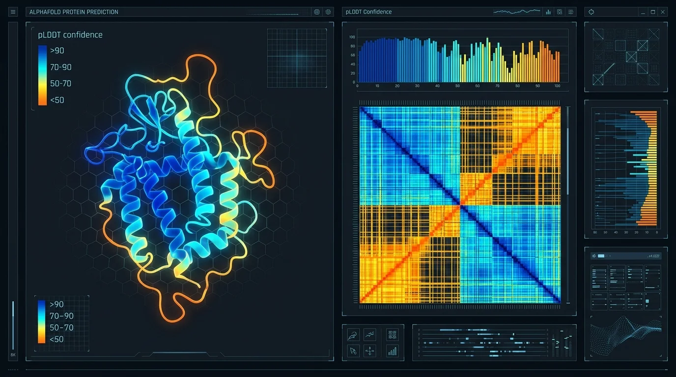 Figure: Residue-level confidence (pLDDT) and inter-domain alignment error (PAE) matrix.
1. pLDDT (predicted Local Distance Difference Test)
A localized per-residue confidence metric bounded between $[0, 100]$:
- $\text{pLDDT} > 90$ (Dark Blue): High atomic precision. Both backbone and side-chain rotamers are highly accurate; suitable for drug docking and enzymatic catalytic pocket modeling.
- $70 < \text{pLDDT} \le 90$ (Light Blue): Reliable backbone topology. Minor side-chain rotamer flexibility may exist.
- $50 < \text{pLDDT} \le 70$ (Yellow): Low confidence; general secondary structure elements may be accurate, but flexible loop regions are uncertain.
- $\text{pLDDT} \le 50$ (Orange / Red): Very low confidence.
Bioengineering Note — Intrinsically Disordered Regions (IDRs): A ribbon model displaying $\text{pLDDT} < 50$ with unstructured, spaghetti-like conformations is often not an algorithmic failure. Roughly 30% of eukaryotic proteomes comprise Intrinsically Disordered Regions (IDRs) that do not adopt a single static 3D structure in isolation, but fold conditionally upon binding a partner or participating in liquid-liquid phase separation (LLPS).
2. PAE (Predicted Aligned Error)
While pLDDT reflects local precision, the PAE matrix ($N_{\text{res}} \times N_{\text{res}}$) quantifies global domain orientation error in angströms ($\text{\AA}$).
- Dark Green Blocks on Diagonal ($0-5\text{ \AA}$): Compact, well-folded structural domains.
- Off-Diagonal Features:
- If inter-domain regions are dark green, the relative orientation between the two domains is rigid and validated.
- If the domains themselves are dark green but their cross-intersection is light green/white ($> 25\text{ \AA}$ PAE), both domains are folded correctly internally but are connected by a flexible linker with no fixed relative orientation.
6. Biology’s “Transistor Moment”: From Silicon Fabs to Biofoundries
In our earlier analysis, “Transistor Wars”, we outlined how semiconductors transformed modern civilization across three pillars. The exact same architectural evolution is now recurring in bioengineering:
1. The Dimensional Leap: Planar to GAAFET vs. 1D Sequence to 3D/4D Complexes
To eliminate leakage currents at the atomic scale, transistors evolved from 2D planar channels to 3D FinFETs and 4D surrounding-gate GAAFET nanosheets. Biology underwent an identical dimensional shift: moving from 1D genomics (reading sequences) to 3D structural proteomics with AlphaFold 2, and into multi-entity atomistic complexes with AlphaFold 3.
2. The Biological Moore’s Law
Gordon Moore’s economic engine drove computing into everyday life by collapsing the cost per transistor. AlphaFold sparked an equivalent cost collapse in biology:
- Pre-AlphaFold: ~200,000 empirical structures solved over 50 years at billions of dollars of cumulative research spending.
- Post-AlphaFold: Over 214 million predicted structures cataloged in the open-access AlphaFold Protein Structure Database (AFDB), covering virtually every known organism. The marginal cost of obtaining a 3D structural hypothesis collapsed to minutes of GPU runtime.
3. The “Fabless” Bioengineering Revolution
TSMC’s pure-play foundry model unbundled chip design from manufacturing, allowing fabless architects (Apple, Nvidia) to innovate without multibillion-dollar capital expenditure.
Generative biological models (RFdiffusion, ProteinMPNN, ESM3) are establishing the exact same fabless model:
- Inverse Folding & De Novo Design: Instead of simply predicting how natural sequences fold ($1\text{D} \rightarrow 3\text{D}$), we invert the pipeline: We sketch a desired catalytic pocket or viral neutralizing epitope in 3D CAD space, generate a de novo backbone via diffusion, and compute an amino acid sequence that will fold into that shape ($3\text{D Function} \rightarrow 1\text{D Sequence}$).
- Designers vs. Manufacturers: Biotech startups no longer screen millions of random variants in wet labs. They computationally filter designs through AlphaFold/PAE filters and outsource physical gene synthesis and robotic expression testing to automated Biofoundries.
7. Conclusion: From Reading to Writing
When Bell Labs demonstrated the first point-contact transistor in 1947, few anticipated that it would evolve into multi-billion-transistor neural processors within decades. With AlphaFold, bioengineering stands at that exact “1947 moment.”
Arthur C. Clarke famously wrote that “any sufficiently advanced technology is indistinguishable from magic.” Yet an engineer’s duty is not to be enchanted by magic, but to understand its mathematics, limits, and tolerances. AlphaFold has not replaced wet-lab experiments; it has elevated structural biologists from structural cartographers to functional molecular architects.
Algorithms compute probability spaces; it is human ingenuity and engineering will that translate those computations into molecules that heal living systems. The era of merely reading biology is over; the era of writing and architecting biology at the atomic scale has begun.
Author: Kerem Uysal — Founder, Tech&Lab
Contact: info@keremuysal.com | info@techandlab.com